Brain Scans of Some Schizophrenics Show Which of the Following
Functional neuroimaging scans such as PET and functional magnetic resonance imaging fMRI taken while individuals perform cognitive tasks particularly those involving memory and. Schizophrenia is a chronic psychiatric disorder.
Schizophrenia What Is Schizophrenia What Are Neurotransmitters How Common Is Schizophrenia What Causes Schizophrenia
Using brain scans researchers have found that many people w schizophrenia have enlarged ventricles brain cavities that contain CSF patients who have enlarged ventricles display more negative symptoms fewer positive symptoms and tend to be more poorly adjusted socially before the onset of schizophrenia have more cognitive disturbances and respond less well to.

. Similarly neuroimaging studies of cognitive remediation in schizophrenia focus differentially on structural functional and connectivity changes. This finding of enlarged ventricles is still the most consistent and reproducible finding in neuroimaging studies of schizophrenia McCarley et al 1999. SYNFIN scanning using IP fragments was created to avoid false positives generated by other scans because of.
Symptoms first appear in. Compare and contrast MRIs PET scans and fMRI. This region is known for producing dopamine and regulates coordinated movement motivation and the reward pathway.
Recruiting broader cortical areas to perform identical tasks. Moreover the researchers found that among schizophrenics those with a busier CA1 had worse delusions. For example the prefrontal cortex is connected to another brain area affected in schizophrenia called the basal ganglia.
The variety of symptoms seen with schizophrenia may be a result of how highly interconnected the brain is. A loss or a decrease in the ability to initiate plans. CT and MRI The majority of the findings seen with CT scan can also be observed with an MRI.
Hallucinations such as hearing voices paranoid delusions and exaggerated or distorted perceptions beliefs and behaviours in affected people. A shrunken cerebral tissue B a smaller than normal thalamus C less activity in the frontal lobes D all the answers are correct. MRIs on schizophrenic brains show the brain regions crucial for regulating motivation emotion perception actions and attention to be abnormal.
These are regions on which episodic memory processing of auditory information and short-term memorydecision making respectively are critically dependent. Scientists have observed abnormalities in the brains of people with schizophrenia when using computed topography CT and magnetic resonance imaging MRI scans. Atrophy loss of brain cellsshrinkage of the hippocampus is among the most notable changes in the brains of people with schizophrenia.
Studies also suggest that the hippocampus a structure in the temporal lobe that influences learning and memory also presents differently in people with schizophrenia. Doctors also believe the brain loses tissue over time. Some findings suggest the presence of brain compensatory mechanisms in schizophrenia eg.
These structural studies have been complemented by functional imaging research which has indicated that schizophrenia is particularly associated with altered function in the prefrontal cingulate and temporal cortex Reference McGuire and Matsumoto McGuire Matsumoto 2004 and that symptoms such as auditory hallucinations and thought disorder are mediated by. Positive psychotic symptoms in this kind of psychological disorder. Psychology questions and answers.
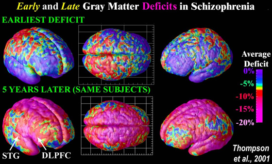
And imaging tools like PET scans and MRIs show that people who have schizophrenia have less gray matter -- the part of the brain that. Ventricular enlargement with cortical atrophy particularly in the frontal lobes is seen in patients with chronic schizophrenia. O shrunken cerebral tissue a smaller than normal thalamus O less activity in the frontal lobes All of these.
Structural brain abnormalities in patients with schizophrenia providing insight into how the condition may develop and respond to treatment have been identified in an internationally. Most patients with a first episode of psychosis will receive a CT brain to rule out organic causes of schizophrenia. In MRI studies of schizophrenia the most consistent findings include reduced gray matter volumes of the medial temporal superior temporal and prefrontal areas.
Question 42 2 pts Brain scans of some schizophrenics show which of the following. The hippocampus the brains learning and memory center is known to be more active in schizophrenics but this study which appears in this months issue of Archives of General Psychiatry pinpoints the part thats hyperactive. Using brain scans researchers have found that many people with schizophrenia have.
Brain scans and postmortem studies show abnormalities in some people with schizophrenia such as enlarged ventricles fluid-filled spaces and reduced size of certain brain regions. CT scans show tightly-packed narrow surface fissuring of the schizophrenic brain. What do brain scans of schizophrenics show.
The following are symptoms of this brain in affected people. It affects how a person behaves thinks feels and interacts with the world and the people around them. On PET scans the activity tends to be abnormally low in the frontal lobes of the schizophrenic brain.
-results consistent with brain damage and dopamine overactivity -CAT scans and MRI scans indicate much larger ventricles in schizophrenics compared to controls which suggest diffuse neuronal damage and cell loss. Reductions in white matter in the frontal lobe before and after diagnosis and treatment an increase in another type of white matter called interstitial white matter neurons below the cortex There are also some inconsistent findings in brain scans on white matter. Brain scans of some people with schizophrenia shows which of the following.
The best diagnosis for schizophrenia is an intensive group of assessments including medical and blood tests and a thorough psychological history and evaluation. Johnstone and colleagues 1976 first demonstrated brain abnormalities on CT scans in patients with schizophrenia. It appears that brain structural change is detectable in both gray and white matter prior to illness onset that active progression of the changes may also begin prior to the onset of clinical symptoms that progressive brain changes may account for the brain structural anomalies seen in chronic schizophrenia and that the structures involved in language.
Brain scans of people with schizophrenia show the following changes to white matter.
Schizophrenia Com Schizophrenia Is A Brain Disease

Evidence That Schizophrenia Is A Brain Disease Schizophrenia
Schizophrenia Com Schizophrenia Is A Brain Disease
No comments for "Brain Scans of Some Schizophrenics Show Which of the Following"
Post a Comment